Tumor Embolization
Advanced Minimally Invasive Treatment for Cancerous and Benign Tumors. Tumor embolization offers a precise and effective way to block a tumor's blood supply. This minimally invasive approach can slow tumor growth, support other treatments, and can be used before surgical resection. Tumors require a good supply of blood from blood vessels to grow. Tumor embolization targets those vessels directly, using image-guided techniques to block the blood supply.
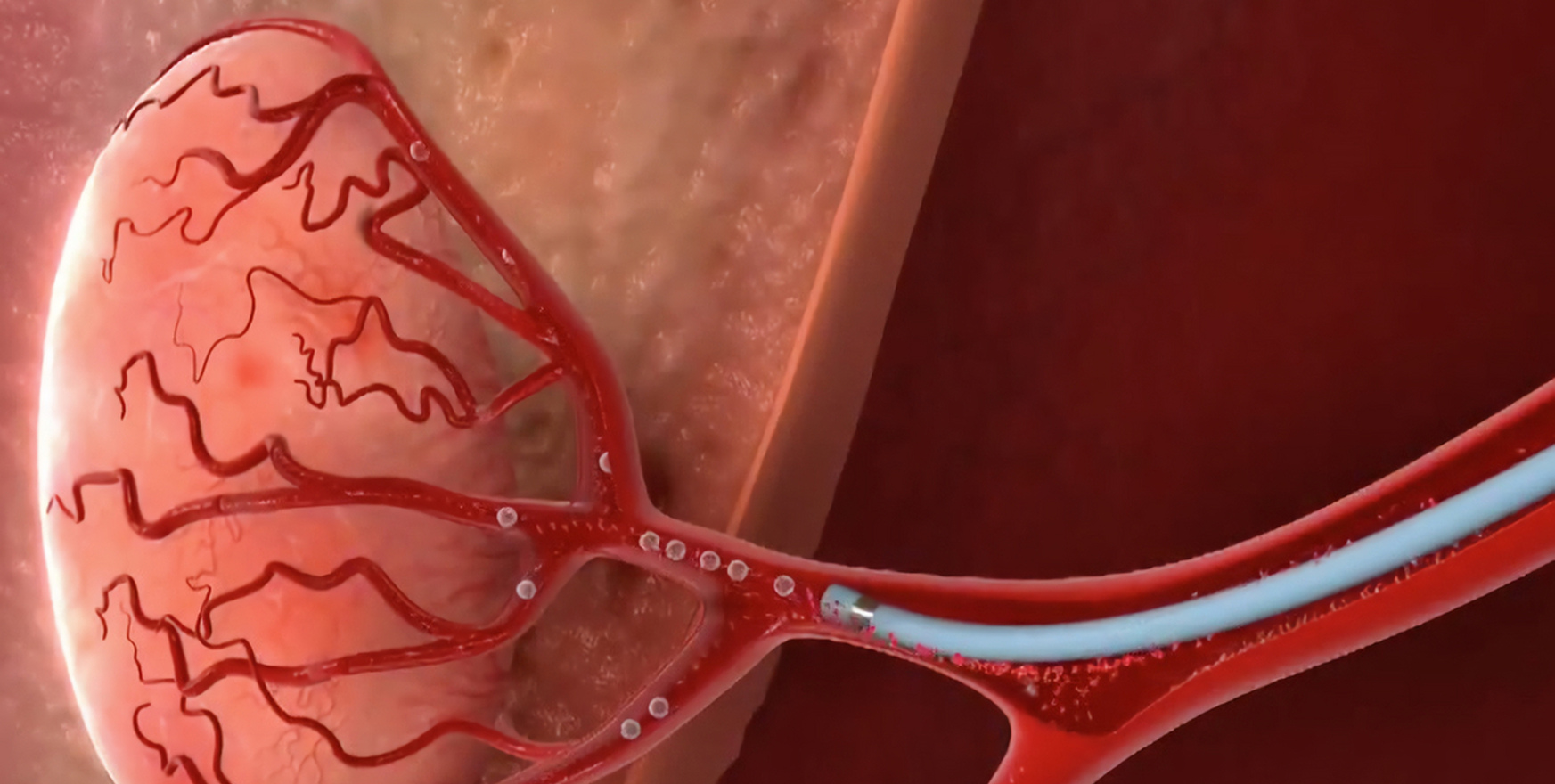
Tumor embolization is a minimally invasive procedure in which a small, thin tube (catheter) is guided into the feeding arteries of a tumor in order either to shut down the blood supply to the tumor or deliver tumor-killing therapy directly to the tumor . Embolization is performed by interventional radiologists and involves using liquid, particles, or microspheres to block blood vessels, redirecting blood flow away from the tumor, causing it to shrink and die.
What is Tumor Embolization?
Embolization involves making a small puncture, typically in the groin or wrist. A catheter is guided through the blood vessels to reach the arteries that feed the tumor. Once in position, the physician releases embolic material, such as beads, foam, coils, or medical glue to block those vessels .
The result is reduced blood flow, less oxygen delivery, and a slower-growing or shrinking tumor. Blocking the tumor's blood supply can also improve the effectiveness of radiation and chemotherapy.
Conditions Treated with Tumor Embolization
Tumor embolization is often used for:
- Liver tumors (primary or metastatic)
- Renal cell carcinoma (kidney cancer)
- Bone tumors
- Uterine fibroids (uterine fibroid embolization)
- Tumors that cause bleeding or pain
- Head, neck, and brain tumors
- Lung tumors
- Highly vascular tumors before surgery
It's also used to reduce blood loss before surgery, making tumor removal safer.
Types of Tumor Embolization
Bland Embolization
Blood Flow Blockage Only
Small beads are injected into the tumor vessel to stop the flow of blood to the tumor without chemotherapy or radiation.
Chemoembolization (TACE)
Chemotherapy + Embolization
Tiny beads infused with chemotherapy are sent through a blood vessel directly into a tumor, delivering high drug concentrations locally.
Radioembolization (TARE)
Radiation + Embolization
Tiny beads with a radioactive isotope (yttrium-90) directly treat tumors with radiation therapy while blocking blood supply.
Preoperative Embolization
Before Surgical Resection
Used before surgery to decrease blood supply to a tumor, reducing intra-operative bleeding and operative times.
Who is a Candidate for Tumor Embolization?
Tumor embolization is typically recommended for patients whose tumors are difficult to remove surgically or when surgery presents high risks due to age, underlying health conditions, or tumor location.
Ideal candidates often include those with:
- Large or highly vascular tumors
- Tumors that bleed easily
- Tumors not responding well to systemic therapies
- Tumors too large for surgical removal
- Patients waiting for liver transplant
- Those needing palliative symptom relief
In some cases, embolization is used preoperatively to reduce bleeding during surgery or to shrink a tumor beforehand.
How Tumor Embolization is Performed
The procedure is performed under local anesthesia with sedation, typically lasting around one hour.
Procedure Steps
Minimally invasive · Image-guided
- The doctor makes a small incision in the groin area to access a blood vessel.
- A catheter is carefully threaded into the vessel.
- Dye is injected so the vessel shows up on imaging.
- Using ultrasound or fluoroscopy (X-ray video), the doctor guides the catheter to the tumor area.
- Medication or agents such as tiny plastic particles, foam, or tiny metal coils are inserted to seal off blood vessels feeding the tumor.
- The interventional radiologist carefully navigates a microcatheter close to the tumor and releases embolic particles.
The particles are usually microspheres less than 0.5 mm in size, which may be combined or loaded with chemotherapy or radiation-emitting substances.
Benefits of Tumor Embolization
Recovery After Tumor Embolization
After tumor embolization, patients may experience mild discomfort, fatigue, or low-grade fever. These symptoms are typical and, in most cases, resolve without issue within a few days.
Post-procedure care includes:
- Laying flat in bed with legs straight for 3 hours (if groin puncture)
- Small bandage over puncture site, removed after 24 hours
- Showering 24 hours after procedure
- Avoiding strenuous activity and lifting over 10lbs for 7 days
- Resuming regular diet and light activity such as walking
- Monitoring for redness, swelling, drainage, or bleeding
You will spend one night in the hospital under close monitoring.
Side Effects and Risks
Common side effects include post-embolization syndrome with fever, nausea, and pain. Less severe side effects include bleeding, bruising, and infection at the puncture site.
- Bleeding or blood vessel injury
- Infection
- Contrast dye reaction
- Allergic reaction to contrast medium
- Non-target embolization (accidental particle release to normal vessels)
- Pain at tumor site
Major complications are rare when performed by operators with appropriate training .
Success Rate of Tumor Embolization
The technical success rate, defined as successful delivery of particles into the tumor, is usually over 95% . Clinical success (partial or complete tumor death and shrinkage) is around 30-50%, though it varies depending on the location, extent, and biology of the underlying disease .
Why Choose Our Tumor Embolization Program?
Our board-certified interventional radiologists specialize in tumor embolization, treating both cancerous and noncancerous tumors using advanced image-guided techniques.
Ready to Consult Dr. Tejendra Ramani?
Book your appointment today and get expert vascular care in Kutch.

Dr. Tejendra Ramani
Consultant Interventional Radiologist
Vascular · Neuro ·
Image-Guided Interventions
MD (Gold Medalist) Neuro & Vascular Interventional Specialist NIMHANS-trained Neuro Intervention, PhD Scholar
View Full Profile